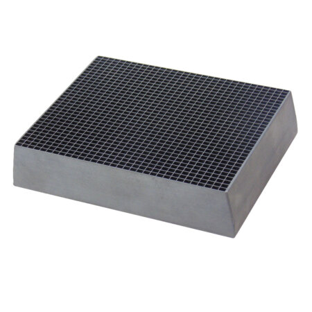
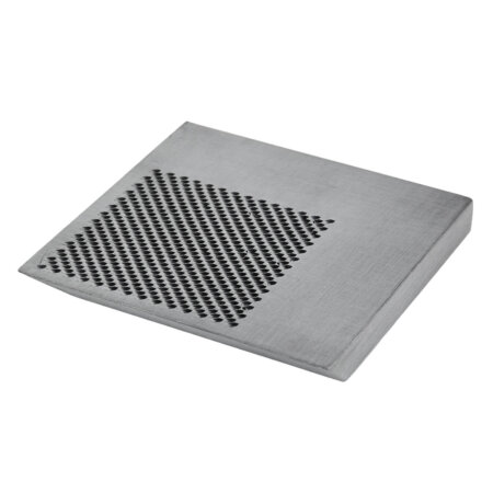
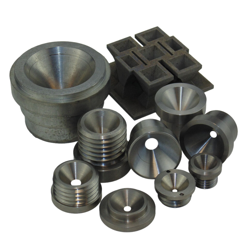
Pinhole Collimators
Pinhole collimators for thyroid scintigraphy, small organ imaging, and preclinical research. Superior spatial resolution with magnification. Single and multi-pinhole configurations available.
Pinhole collimators achieve the highest spatial resolution of any collimator type, using a single small aperture to create magnified, inverted images of small organs and structures. Nuclear Shields manufactures pinhole collimators for clinical thyroid imaging, pediatric applications, and preclinical small-animal research.
Multi-pinhole configurations
The low sensitivity of single-pinhole imaging has driven development of multi-pinhole collimators, particularly for preclinical small-animal SPECT. Multiple pinholes viewing the same source volume increase sensitivity proportionally while maintaining high resolution, at the cost of image overlap (multiplexing) that must be resolved during reconstruction. Nuclear Shields manufactures multi-pinhole collimators for preclinical imaging systems, with aperture configurations optimized for specific detector geometries and target resolutions.

Product description
Single-aperture imaging geometry
Unlike multi-hole collimators that accept photons across many parallel channels, a pinhole collimator uses a single small aperture. Typically 2–6 mm diameter. Through which all detected photons must pass. This geometry operates similarly to a camera obscura or pinhole camera, producing an inverted image on the detector.
The small aperture provides exceptional angular selectivity, accepting only photons from a narrow cone of directions. This geometric constraint delivers spatial resolution superior to parallel hole collimators, limited primarily by the pinhole diameter rather than hole length and septal penetration.
Magnification and resolution
Pinhole collimators produce magnified images when the source is closer to the aperture than the aperture-to-detector distance. This magnification effectively reduces the contribution of intrinsic detector resolution to overall system resolution, further improving spatial detail.
Resolution improves as: – Pinhole diameter decreases – Source-to-aperture distance decreases – Magnification increases
However, sensitivity drops dramatically with smaller pinholes and increased source distance, creating the fundamental resolution-sensitivity trade-off. Pinhole imaging requires the source to be positioned close to the aperture, making this collimator type practical only for small, superficial organs.
Contact usAdditional images
Product attachments
Login to see other downloads, otherwise contact us